What is the Shift-and-Persist Model?

Introduction

The Shift-and-persist model has emerged in order to account for unintuitive, positive health outcomes in some individuals of low socioeconomic status.

A large body of research has previously linked low socioeconomic status to poor physical and mental health outcomes, including early mortality. Low socioeconomic status is hypothesized to get “under the skin” by producing chronic activation of the sympathetic nervous system and hypothalamic–pituitary–adrenal axis, which increases allostatic load, leading to the pathogenesis of chronic disease. However, some individuals of low socioeconomic status do not appear to experience the expected, negative health effects associated with growing up in poverty. To account for this, the Shift-and-Persist Model proposes that, as children, some individuals of low socioeconomic status learn adaptive strategies for regulating their emotions (“shifting”) and focusing on their goals (“persisting”) in the face of chronic adversity. According to this model, the use of shift-and-persist strategies diminishes the typical negative effects of adversity on health by leading to more adaptive biological, cognitive, and behavioural responses to daily stressors.

Shift Strategies

Broadly, “shift” strategies encompass a variety of cognitive and emotion self-regulation approaches that individuals use to deal with stress, including cognitive restructuring, reframing, reappraisal, and acceptance strategies, which change the meaning of a stressor or reduce its emotional impact. These shift strategies particularly focus on changing one’s response to a stressor, instead of attempting to change the situation or stressor itself. As shift strategies depend more on internal processes (self-control and regulation), than external resources, it is hypothesized that shift strategies may be particularly adaptive responses to the chronic, uncontrollable stressors that are associated with low socioeconomic status.

Persist Strategies

According to Chen and Miller, “persist” strategies are any strategies that help individuals to maintain optimism about the future, create meaning from their experiences of challenge and hardship, and persist “with strength in the face of adversity.”

Measurement

To evaluate the combination of shift-and-persist strategy use, distinct “shift” and “persist” constructs were initially measured separately by using multiple, self-report measures of reappraisal, emotional reactivity, and future orientation in early research on this model.

In 2015, Chen and colleagues published the Shift-and-Persist Scale, which is a combined self-report measure that assesses both shift and persist strategies. The Shift-and-Persist Scale has been validated for use with adults and teenagers. The questionnaire asks respondents to rate how well 14 statements about various approaches to dealing with life stressors apply to them on a 1-4 scale. Out of the 14 items on the measure, 4 assess a respondent’s use of shift strategies, 4 load onto persist strategies, and 6 items are non-relevant distractors that are ignored during scoring. When scoring the Shift-and-Persist Scale, one item (#4) is reverse-scored. This scale is publicly available online.

A simplified 5-item Shift-and-Persist scale has also been published for use with younger children and adolescents (ages 9–15). Total scores on this version of the Shift-and-Persist Scale range from 0-20, such that higher scores are indicative of greater use of shift-and-persist strategies. This scale is also publicly available online and has been previously used in research with children from kindergarten through 8th grade.

Proposed Mechanisms

Reduction of the Harmful Biological Effects of Stress

The shift-and-persist model mainly hypothesizes that these strategies have protective effects for the health of low socioeconomic status individuals because they affect biological and physiological stress response tendencies that are relevant for disease. There is some evidence that shift responses (e.g. reappraisal) to acute stressors are associated with attenuated physiological responses to stress, including reduced cardiovascular reactivity. Specifically, reappraisal has been linked to a “healthier” pattern of hypothalamic–pituitary–adrenal axis response characterised by a rapid return to homeostasis (i.e., faster cortisol recovery) in the wake of a stressor. Persist tendencies, such as optimism, have also been associated with adaptive immune responses and faster cortisol recovery. By constraining the magnitude and duration of biological stress responses, including cardiovascular, hypothalamic–pituitary–adrenal axis, and inflammatory responses to stress, shift-and-persist responses are hypothesized to prevent the wear and tear on these systems that increases allostatic load and risk for chronic diseases of aging.

Cross-sectional studies provide some evidence that greater emotion regulation abilities are associated with reduced health risk on a variety of indicators of allostatic load. Similarly, self-reported trait levels of optimism and purpose in life have been linked to better concurrent health and health trajectories over time. However, most of the health benefits associated with shift-and-persist consistent strategies are only seen in low socioeconomic status samples.

Enhancement of Adaptive Biological Stress-Recovery Systems

Another alternative, but not mutually exclusive hypothesis, is that shift-and-persist strategies affect health by increasing or up-regulating biological responses that enhance stress recovery and resilience. In particular, the parasympathetic nervous system’s functioning may be enhanced by shift-and-persist response tendencies. Emotion regulation abilities that are consistent with shift-coping have been linked to greater parasympathetic nervous system functioning at rest, as indexed by higher levels of high-frequency heart rate variability. Further, the parasympathetic nervous system is highly integrated with, and may contribute to the down-regulation of hypothalamic–pituitary–adrenal axis and immune system stress responses that influence allostatic load over time. Although parasympathetic nervous system activity is correlated with aspects of shift-and-persist coping, it is not yet established that the use of these strategies actually increases parasympathetic nervous system activity.

The oxytocin system has also been identified as another potential mechanism by which shift-and-persist strategies could influence health outcomes. Oxytocin is a hormone that has been linked to a wide range of positive social and emotional functions and can be used to effectively attenuate hypothalamic–pituitary–adrenal axis and sympathetic nervous system responses to stress. However, there is little research examining the interplay between shift-and-persist strategy use and the oxytocin system.

Impact on Health Behaviours

It has also been proposed that shift-and-persist strategies may buffer health outcomes in individuals of low socioeconomic status by affecting health behaviours. Previous research has demonstrated that, regardless of socioeconomic status, individuals with emotion regulation difficulties are also likely to engage in poorer health behaviours, including over-eating, sedentary lifestyle, risky sexual health behaviours, and drug use. Individuals of low socioeconomic status who learn to regulate their emotions more effectively, by using “shift” strategies in childhood, may be more likely than their peers with emotion regulation difficulties to establish and sustain positive health behaviours throughout development. Similarly, persist strategies that help individuals to maintain a positive focus on the future may also affect wellbeing through health behaviours. Prior studies have linked being “future-oriented” to lower levels of drug use and sexual risk behaviours. Therefore, it is possible that individuals who regularly use shift-and-persist strategies will be more likely to practice positive health behaviours, which promote healthy development and aging.

However, it is important to note that the relationships between emotion regulation abilities and health behaviour are bidirectional. Health behaviours, such as physical activity and sleep hygiene, can also have powerful effects on our capacity to successfully regulate emotions.

Research Support for Associations with Health

Since 2012, integrative research groups concerned with clinical health psychology, social psychology, psychoneuroimmunology, and public health have begun to evaluate the relationships postulated by the shift-and-persist model. The majority of empirical studies on this topic test whether shift-and-persist strategies are associated with differential health outcomes in low vs. high socioeconomic status samples.

Thus far, high levels of shift-and-persist strategy use have been linked to:

  • Lower total allostatic load in adults who grew up in low, but not high, socioeconomic status households.
  • Lower body mass index in children from low, but not high, socioeconomic status families.
  • Reduced low-grade inflammation in adolescents (and parents) from low socioeconomic status families.
  • A “healthier” profile of hypothalamic–pituitary–adrenal axis functioning, as indexed by diurnal cortisol in children from low socioeconomic status families.
  • Lower levels of asthma-related impairment and inflammation in children from low, but not high, socioeconomic status families.
  • Better asthma profiles in children and teens from families reporting low, but not high, perceived social status.
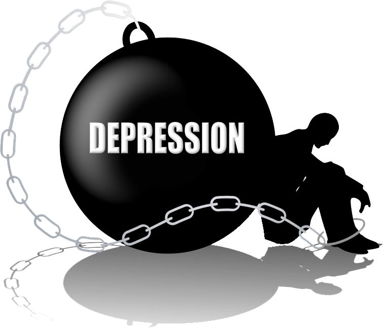
  • Lower levels of depressive symptoms in Latinx youth from low, but not high, income families.

Although it has been proposed that a variety of psychological interventions for at-risk youth of low socioeconomic status may reduce health disparities, in part, by increasing shift-and-persist tendencies in families, the majority of studies on shift-and-persist have been cross-sectional. Therefore, it remains unknown if shift-and-persist strategies play a causal role in reducing the negative impact of low socioeconomic status on health. More longitudinal and treatment studies are needed to evaluate directional and causal hypotheses based upon the shift-and-persist model.

This page is based on the copyrighted Wikipedia article < https://en.wikipedia.org/wiki/Shift-and-persist_model >; it is used under the Creative Commons Attribution-ShareAlike 3.0 Unported License (CC-BY-SA). You may redistribute it, verbatim or modified, providing that you comply with the terms of the CC-BY-SA.

What is Allostatic Load?

Introduction

Allostatic load is “the wear and tear on the body” which accumulates as an individual is exposed to repeated or chronic stress.

The term was coined by Bruce McEwen and Stellar in 1993. It represents the physiological consequences of chronic exposure to fluctuating or heightened neural or neuroendocrine response which results from repeated or prolonged chronic stress.

Regulatory Model

The term allostatic load is “the wear and tear on the body” which accumulates as an individual is exposed to repeated or chronic stress. It was coined by McEwen and Stellar in 1993.

The term is part of the regulatory model of allostasis, where the predictive regulation or stabilisation of internal sensations in response to stimuli is ascribed to the brain. Allostasis involves the regulation of homeostasis in the body to decrease physiological consequences on the body. Predictive regulation refers to the brain’s ability to anticipate needs and prepare to fulfil them before they arise.

Part of efficient regulation is the reduction of uncertainty. Humans naturally do not like feeling as if surprise is inevitable. Because of this, we constantly strive to reduce the uncertainty of future outcomes, and allostasis helps us do this by anticipating needs and planning how to satisfy them ahead of time. But it takes a considerable amount of the brain’s energy to do this, and if it fails to resolve the uncertainty, the situation may become chronic and result in the accumulation of allostatic load.

The concept of allostatic load provides that:

“the neuroendocrine, cardiovascular, neuroenergetic, and emotional responses become persistently activated so that blood flow turbulences in the coronary and cerebral arteries, high blood pressure, atherogenesis, cognitive dysfunction and depressed mood accelerate disease progression.”

All long-standing effects of continuously activated stress responses are referred to as allostatic load. Allostatic load can result in permanently altered brain architecture and systemic pathophysiology.

Allostatic load minimises an organism’s ability to cope with and reduce uncertainty in the future.

Types

McEwen and Wingfield propose two types of allostatic load with different aetiologies and distinct consequences:

  • Type 1 allostatic load occurs when energy demand exceeds supply, resulting in activation of the emergency life history stage. This serves to direct the animal away from normal life history stages into a survival mode that decreases allostatic load and regains positive energy balance. The normal life cycle can be resumed when the perturbation has passed. Typical situations ending up in type 1 allostasis are starvation, hibernation and critical illness. Of note, the life-threatening consequences of critical illness may be both cause and consequences of allostatic load.
  • Type 2 allostatic load results from sufficient or even excess energy consumption being accompanied by social conflict or other types of social dysfunction. The latter is the case in human society and certain situations affecting animals in captivity. In all cases, secretion of glucocorticosteroids and activity of other mediators of allostasis such as the autonomic nervous system, CNS neurotransmitters, and inflammatory cytokines wax and wane with allostatic load. If allostatic load is chronically high, then pathologies may develop. Type 2 allostatic overload does not trigger an escape response, and can only be counteracted through learning and changes in the social structure.

Whereas both types of allostatic load are associated with increased release of cortisol and catecholamines, they differentially affect thyroid homeostasis: Concentrations of the thyroid hormone triiodothyronine are decreased in type 1 allostasis, but elevated in type 2 allostasis. This may result from an interaction of type 2 allostatic load with the set point of thyroid function.

Measurement

Allostatic load is generally measured through a composite index of indicators of cumulative strain on several organs and tissues, primarily biomarkers associated with the neuroendocrine, cardiovascular, immune and metabolic systems.

Indices of allostatic load are diverse across studies and are frequently assessed differently, using different biomarkers and different methods of assembling an allostatic load index. Allostatic load is not unique to humans and may be used to evaluate the physiological effects of chronic or frequent stress in non-human primates as well.

In the endocrine system, the increase or repeated levels of stress results in the increased levels of the hormone Corticotropin-Releasing Factor (CRH), which is associated with activation of hypothalamic-pituitary-adrenal (HPA) axis. The HPA axis is the central stress response system responsible for modulating inflammatory responses throughout the body. Prolonged stress levels can lead to decreased levels of cortisol in the morning and increased levels in the afternoon, leading to greater daily output of cortisol which in the long term increases blood sugar levels.

In the nervous system, structural and functional abnormalities are a result of chronic prolonged stress. The increase of stress levels causes a shortening of dendrites in a neuron. Therefore, the shortening of dendrites causes the decrease in attention. Chronic stress also causes greater response to fear of the unlearned in the nervous system, and fear conditioning.

In the immune system, the increase in levels of chronic stress results in the elevation of inflammation. The increase in inflammation levels is caused by the ongoing activation of the sympathetic nervous system. The impairment of cell-mediated acquired immunity is also a factor resulting in the immune system due to chronic stress.

Relationship to Allostasis and Homeostasis

The largest contribution to the allostatic load is the effect of stress on the brain. Allostasis is the system which helps to achieve homeostasis. Homeostasis is the regulation of physiological processes, whereby systems in the body respond to the state of the body and to the external environment. The relationship between allostasis and allostatic load is the concept of anticipation. Anticipation can drive the output of mediators. Examples of mediators include hormones and cortisol. Excess amounts of such mediators will result in an increase in allostatic load, contributing to anxiety and anticipation.

Allostasis and allostatic load are related to the amount of health-promoting and health-damaging behaviours like for example cigarette smoking, consumption of alcohol, poor diet and physical inactivity.

Three physiological processes cause an increase in allostatic load:

  • Frequent stress: the magnitude and frequency of response to stress is what determines the level of allostatic load which affects the body.
  • Failed shut-down: the inability of the body to shut off while stress accelerates and levels in the body exceed normal levels, for example, elevated blood pressure.
  • Inadequate response: the failure of the body systems to respond to challenge, for example, excess levels of inflammation due to inadequate endogenous glucocorticoid responses.

The importance of homeostasis is to regulate the stress levels encountered on the body to reduce allostatic load.

Dysfunctional allostasis causes allostatic load to increase which may, over time, lead to disease, sometimes with decompensation of the allostatically controlled problem. Allostatic load effects can be measured in the body. When tabulated in the form of allostatic load indices using sophisticated analytical methods, it gives an indication of cumulative lifetime effects of all types of stress on the body.

Causes of Allostatic Load

Type 1 allostatic load represents the adaptive response to an absolute lack in energy, glutathione and several macronutrients. It also includes predictive responses, e.g. in hibernation, infection and depression.

Type 2 allostatic load results from an expected mismatch of energy demand and supply. It is triggered by psychosocial stress, e.g. due to low socioeconomic status, major life events and environmental stressors. This association explains the increased risk for cardiovascular disease and chronic conditions like obesity, diabetes, hypertension and psychotic conditions in subjects that were exposed to psychosocial trauma, social disadvantage and discrimination. Socio-cultural mechanisms tend to augment this relation by perpetuating disparity even in the quality of health care, which tends to be inferior in socially disadvantaged population strata.

Implications of Allostatic Load on Health

Increased allostatic load constitutes a significant health hazard. Several studies documented a strong association of allostatic load to the incidence of coronary heart disease, to surrogate markers of cardiovascular health and to hard endpoints, including cause-specific and all-cause mortality. Mediators connecting allostatic load to morbidity and mortality include the function of the autonomic nervous system, cytokines and stress hormones, e.g. catecholamines, cortisol and thyroid hormones.

Reducing Risk

To reduce and manage high allostatic load, an individual should pay attention to structural and behavioural factors. Structural factors include the social environment, and access to health services. Behavioural factors include diet, physical health and tobacco smoking, which can lead to chronic disease. Actions such as tobacco smoking are brought about from the stress levels that an individual experiences. Therefore, controlling stress levels from the beginning, for example by not leading to tobacco smoking, will reduce the chance of chronic disease development and high allostatic load.

Low socio-economic status (SES) affects allostatic load and therefore, focusing on the causes of low SES will reduce allostatic load levels. Reducing societal polarisation, material deprivation, and psychological demands on health helps to manage allostatic load. Support from the community and the social environment can manage high allostatic load. In addition, healthy lifestyle that encompasses a broad array of lifestyle change including healthy eating and regular physical exercise may reduce allostatic load. Empowering financial help from the government allows people to gain control and improve their psychological health. Improving inequalities in health decreases the stress levels and improves health by reducing high allostatic load on the body.

Interventions can include encouraging sleep quality and quantity, social support, self-esteem and wellbeing, improving diet, avoiding alcohol or drug consumption and participating in physical activity. Providing cleaner and safer environments and the incentive towards a higher education will reduce the chance of stress and improve mental health significantly, therefore, reducing the onset of high allostatic load.

Allostatic load differs by sex and age, and the social status of an individual. Protective factors could, at various times of an individual’s life span, be implemented to reduce stress and, in the long run, eliminate the onset of allostatic load. Protective factors include parental bonding, education, social support, healthy workplaces, a sense of meaning towards life and choices being made, and positive feelings in general.

What is the Gut-Brain Axis?

Introduction

The gut-brain axis is the biochemical signalling that takes place between the gastrointestinal tract (GI tract) and the central nervous system (CNS).

The term “gut-brain axis” is occasionally used to refer to the role of the gut flora in the interplay as well, whereas the term “microbiota–gut–brain (MGB or BGM) axis” explicitly includes the role of gut flora in the biochemical signalling events that take place between the GI tract and CNS.

Broadly defined, the gut-brain axis includes the central nervous system, neuroendocrine and neuroimmune systems, including the hypothalamic-pituitary-adrenal axis (HPA axis), sympathetic and parasympathetic arms of the autonomic nervous system, including the enteric nervous system and the vagus nerve, and the gut microbiota. The first of the brain-gut interactions shown, was the cephalic phase of digestion, in the release of gastric and pancreatic secretions in response to sensory signals, such as the smell and sight of food. This was first demonstrated by Pavlov.

Interest in the field was sparked by a 2004 study showing that germ-free (GF) mice showed an exaggerated HPA axis response to stress compared to non-GF laboratory mice.

As of October 2016, most of the work done on the role of gut flora in the gut-brain axis had been conducted in animals, or on characterising the various neuroactive compounds that gut flora can produce. Studies with humans – measuring variations in gut flora between people with various psychiatric and neurological conditions or when stressed, or measuring effects of various probiotics (dubbed “psychobiotics” in this context) – had generally been small and were just beginning to be generalised. Whether changes to gut flora are a result of disease, a cause of disease, or both in any number of possible feedback loops in the gut–brain axis, remained unclear.

Gut Flora

The gut flora is the complex community of microorganisms that live in the digestive tracts of humans and other animals. The gut metagenome is the aggregate of all the genomes of gut microbiota. The gut is one niche that human microbiota inhabit.

In humans, the gut microbiota has the largest quantity of bacteria and the greatest number of species, compared to other areas of the body. In humans, the gut flora is established at one to two years after birth; by that time, the intestinal epithelium and the intestinal mucosal barrier that it secretes have co-developed in a way that is tolerant to, and even supportive of, the gut flora and that also provides a barrier to pathogenic organisms.

The relationship between gut flora and humans is not merely commensal (a non-harmful coexistence), but rather a mutualistic relationship. Human gut microorganisms benefit the host by collecting the energy from the fermentation of undigested carbohydrates and the subsequent absorption of short-chain fatty acids (SCFAs), acetate, butyrate, and propionate. Intestinal bacteria also play a role in synthesizing vitamin B and vitamin K as well as metabolising bile acids, sterols, and xenobiotics. The systemic importance of the SCFAs and other compounds they produce are like hormones and the gut flora itself appears to function like an endocrine organ; dysregulation of the gut flora has been correlated with a host of inflammatory and autoimmune conditions.

The composition of human gut flora changes over time, when the diet changes, and as overall health changes.

Enteric Nervous System

The enteric nervous system is one of the main divisions of the nervous system and consists of a mesh-like system of neurons that governs the function of the gastrointestinal system; it has been described as a “second brain” for several reasons. The enteric nervous system can operate autonomously. It normally communicates with the central nervous system (CNS) through the parasympathetic (e.g. via the vagus nerve) and sympathetic (e.g. via the prevertebral ganglia) nervous systems. However, vertebrate studies show that when the vagus nerve is severed, the enteric nervous system continues to function.

In vertebrates, the enteric nervous system includes efferent neurons, afferent neurons, and interneurons, all of which make the enteric nervous system capable of carrying reflexes in the absence of CNS input. The sensory neurons report on mechanical and chemical conditions. Through intestinal muscles, the motor neurons control peristalsis and churning of intestinal contents. Other neurons control the secretion of enzymes. The enteric nervous system also makes use of more than 30 neurotransmitters, most of which are identical to the ones found in CNS, such as acetylcholine, dopamine, and serotonin. More than 90% of the body’s serotonin lies in the gut, as well as about 50% of the body’s dopamine; the dual function of these neurotransmitters is an active part of gut-brain research.

The first of the gut-brain interactions was shown to be between the sight and smell of food and the release of gastric secretions, known as the cephalic phase, or cephalic response of digestion.

Gut-Brain Integration

The gut-brain axis, a bidirectional neurohumoral communication system, is important for maintaining homeostasis and is regulated through the central and enteric nervous systems and the neural, endocrine, immune, and metabolic pathways, and especially including the hypothalamic-pituitary-adrenal axis (HPA axis). That term has been expanded to include the role of the gut flora as part of the “microbiome-gut-brain axis”, a linkage of functions including the gut flora.

Interest in the field was sparked by a 2004 study (Nobuyuki Sudo and Yoichi Chida) showing that germ-free mice (genetically homogeneous laboratory mice, birthed and raised in an antiseptic environment) showed an exaggerated HPA axis response to stress, compared to non-GF laboratory mice.

The gut flora can produce a range of neuroactive molecules, such as acetylcholine, catecholamines, γ-aminobutyric acid, histamine, melatonin, and serotonin, which are essential for regulating peristalsis and sensation in the gut. Changes in the composition of the gut flora due to diet, drugs, or disease correlate with changes in levels of circulating cytokines, some of which can affect brain function. The gut flora also release molecules that can directly activate the vagus nerve, which transmits information about the state of the intestines to the brain.

Likewise, chronic or acutely stressful situations activate the hypothalamic-pituitary-adrenal axis, causing changes in the gut flora and intestinal epithelium, and possibly having systemic effects. Additionally, the cholinergic anti-inflammatory pathway, signalling through the vagus nerve, affects the gut epithelium and flora. Hunger and satiety are integrated in the brain, and the presence or absence of food in the gut and types of food present also affect the composition and activity of gut flora.

That said, most of the work that has been done on the role of gut flora in the gut-brain axis has been conducted in animals, including the highly artificial germ-free mice. As of 2016, studies with humans measuring changes to gut flora in response to stress, or measuring effects of various probiotics, have generally been small and cannot be generalised; whether changes to gut flora are a result of disease, a cause of disease, or both in any number of possible feedback loops in the gut-brain axis, remains unclear.

The history of ideas about a relationship between the gut and the mind dates from the nineteenth century. The concepts of dyspepsia and neurasthenia gastrica referred to the influence of the gut on human emotions and thoughts.

Gut-Brain-Skin Axis

A unifying theory that tied gastrointestinal mechanisms to anxiety, depression, and skin conditions such as acne was proposed as early as 1930. In a paper in 1930, it was proposed that emotional states might alter normal intestinal flora which could lead to increased intestinal permeability and therefore contribute to systemic inflammation. Many aspects of this theory have been validated since then. Gut microbiota and oral probiotics have been found to influence systemic inflammation, oxidative stress, glycaemic control, tissue lipid content, and mood.

Research

Probiotics

A 2016 systematic review of laboratory animal studies and preliminary human clinical trials using commercially available strains of probiotic bacteria found that certain species of the Bifidobacterium and Lactobacillus genera (i.e. B. longum, B. breve, B. infantis, L. helveticus, L. rhamnosus, L. plantarum, and L. casei) had the most potential to be useful for certain central nervous system disorders.

Anxiety and Mood Disorders

As of 2018 work on the relationship between gut flora and anxiety disorders and mood disorders, as well as attempts to influence that relationship using probiotics or prebiotics (called “psychobiotics”), was at an early stage, with insufficient evidence to draw conclusions about a causal role for gut flora changes in these conditions, or about the efficacy of any probiotic or prebiotic treatment.

People with anxiety and mood disorders tend to have gastrointestinal problems; small studies have been conducted to compare the gut flora of people with major depressive disorder and healthy people, but those studies have had contradictory results.

Much interest was generated in the potential role of gut flora in anxiety disorders, and more generally in the role of gut flora in the gut-brain axis, by studies published in 2004 showing that germ-free mice have an exaggerated HPA axis response to stress caused by being restrained, which was reversed by colonising their gut with a Bifidobacterium species. Studies looking at maternal separation for rats shows neonatal stress leads to long-term changes in the gut microbiota such as its diversity and composition, which also led to stress and anxiety-like behaviour. Additionally, while much work had been done as of 2016 to characterise various neurotransmitters known to be involved in anxiety and mood disorders that gut flora can produce (for example, Escherichia, Bacillus, and Saccharomyces species can produce noradrenalin; Candida, Streptococcus, and Escherichia species can produce serotonin, etc.) the interrelationships and pathways by which the gut flora might affect anxiety in humans were unclear.

In one study, germ-free mice underwent faecal transplants with microbes from humans with or without major depressive disorder (MDD). Mice with microbes from humans with MDD displayed more behaviours associated with anxiety and depression than mice transplanted with microbes from humans without MDD. The taxonomic composition of microbiota between depressed patients and healthy patients, as well as between the respective mice, also differed. Germ-free mice in another study also displayed behaviours associated with anxiety and depression as compared to mice with normal microbiota, and had higher levels of corticosterone after exposure to behavioural tests. Using rodents in microbiome and mental health studies allows researchers to compare behaviour and microbial composition of rodents to humans, ideally to elucidate therapeutic application for mental disorders.

Additionally, there is a link between the gut microbiome, mood disorders and anxiety, and sleep. The microbial composition of the gut microbiome changes depending on the time of day, meaning that throughout the day, the gut is exposed to varying metabolites produced by the microbes active during that time. These time-dependent microbial changes are associated with differences in the transcription of circadian clock genes involved in circadian rhythm. One mouse study showed that altering clock gene transcription by disrupting circadian rhythm, such as through sleep deprivation, potentially has a direct effect on the composition of the gut microbiome. Another study found that mice that could not produce the CLOCK protein, made by a clock gene, were more likely to develop depression. Stress and sleep disturbances can lead to greater gut mucosal permeability via activation of the HPA axis. This in turn causes immune inflammatory responses that contribute to the development of illnesses that cause depression and anxiety.

Autism

Around 70% of people with autism also have gastrointestinal problems, and autism is often diagnosed at the time that the gut flora becomes established, indicating that there may be a connection between autism and gut flora. Some studies have found differences in the gut flora of children with autism compared with children without autism – most notably elevations in the amount of Clostridium in the stools of children with autism compared with the stools of the children without – but these results have not been consistently replicated. Many of the environmental factors thought to be relevant to the development of autism would also affect the gut flora, leaving open the question of whether specific developments in the gut flora drive the development of autism or whether those developments happen concurrently. As of 2016, studies with probiotics had only been conducted with animals; studies of other dietary changes to treat autism have been inconclusive.

Parkinson’s Disease

As of 2015, one study had been conducted comparing the gut flora of people with Parkinson’s disease to healthy controls; in that study people with Parkinson’s had lower levels of Prevotellaceae and people with Parkinson’s who had higher levels of Enterobacteriaceae had more clinically severe symptoms; the authors of the study drew no conclusions about whether gut flora changes were driving the disease or vice versa.

Major Depressive Disorder: Childhood Trauma

Research Paper Title

Major depressive disorder with childhood trauma: Clinical characteristics, biological mechanism, and therapeutic implications.

Background

Major depressive disorder (MDD) is a main type of mood disorder, characterised by significant and lasting depressed mood.

Until now, the pathogenesis of MDD is not clear, but it is certain that biological, psychological, and social factors are involved.

Childhood trauma is considered to be an important factor in the development of this disease.

Previous studies have found that nearly half of the patients with MDD have experienced childhood trauma, and different types of childhood trauma, gender, and age show different effects on this disease.

In addition, the clinical characteristics of MDD patients with childhood trauma are also different, which often have more severe depressive symptoms, higher risk of suicide, and more severe cognitive impairment.

The response to antidepressants is also worse.

In terms of biological mechanisms and marker characteristics, the serotonin transporter gene and the FKBP prolyl isomerase 5 have been shown to play an important role in MDD and childhood trauma.

Moreover, some brain imaging and biomarkers showed specific features, such as changes in gray matter in the dorsal lateral prefrontal cortex, and abnormal changes in hypothalamic-pituitary-adrenal axis function.

Reference

Guo, W., Liu, J. & Li, L. (2020) Major depressive disorder with childhood trauma:Clinical characteristics, biological mechanism, and therapeutic implications. Zhong nan da xue xue bao. Journal of Central South University. 45(4), pp.462-468. doi: 10.11817/j.issn.1672-7347.2020.190699.

The Effects of Childhood Trauma on Increased Cortisol Levels in Patients with Glucocorticoid Resistance

Research Paper Title

Childhood Trauma, HPA Axis Activity and Antidepressant Response in Patients with Depression.

Background

Childhood trauma is among the most potent contributing risk factors for depression and is associated with poor treatment response.

Hypothalamic-pituitary-adrenal (HPA) axis abnormalities have been linked to both childhood trauma and depression, but the underlying mechanisms are poorly understood.

The present study aimed to investigate the link between childhood trauma, HPA axis activity and antidepressant response in patients with depression.

Methods

As part of the Wellcome Trust NIMA consortium, 163 depressed patients and 55 healthy volunteers were included in this study.

Adult patients meeting Structured Clinical Interview for Diagnostic and Statistical Manual Version-5 criteria for major depression were categorised into subgroups of treatment responder (n=42), treatment non-responder (n=80) and untreated depressed (n=41) based on current depressive symptom severity measured by the 17-item Hamilton Rating Scale for Depression and exposure to antidepressant medications established by Antidepressant Treatment Response Questionnaire. Childhood Trauma Questionnaire was obtained.

Baseline serum C-reactive protein was measured using turbidimetric detection. Salivary cortisol was analysed at multiple time points during the day using the ELISA technique. Glucocorticoid resistance was defined as the coexistence of hypercortisolemia and inflammation.

Results

The results show that treatment non-responder patients had higher exposure to childhood trauma than responders.

No specific HPA axis abnormalities were found in treatment non-responder depressed patients.

Untreated depressed showed increased diurnal cortisol levels compared with patients on antidepressant medication, and higher prevalence of glucocorticoid resistance than medicated patients and controls.

The severity of childhood trauma was associated with increased diurnal cortisol levels only in individuals with glucocorticoid resistance.

Conclusions

The researchers argue their findings suggest that the severity of childhood trauma experience contributes to a lack of response to antidepressant treatment.

The effects of childhood trauma on increased cortisol levels are specifically evident in patients with glucocorticoid resistance and suggest glucocorticoid resistance as a target for the development of personalised treatment for a subgroup of depressed patients with a history of childhood trauma rather than for all patients with resistance to antidepressant treatment.

Reference

Nikkheslat, N., McLaughlin, A.P., Hastings, C., Zajkowska, Z., Nettis, M.A., Mariani, N., Enache, D., Lombardo, G., Pointon, L., Cowen, P.J., Cavanagh, J., Harrison, N.A., Bullmore, E.T., Pariante, C.M., Mondelli, V. & NIMA Consortium. (2019) Childhood Trauma, HPA Axis Activity and Antidepressant Response in Patients with Depression. Brain, Behavior, and Immunity. pii: S0889-1591(19)30702-0. doi: 10.1016/j.bbi.2019.11.024. [Epub ahead of print].