Sequenced Treatment Alternatives to Relieve Depression (STARD) was a collaborative study on the treatment of depression, funded by the National Institute of Mental Health. Its main focus was on the treatment of depression in patients where the first prescribed antidepressant proved inadequate. A key feature of the study was its aim to be more generalisable to real clinical situations; this was done through the use of minimal exclusion criteria, incorporating patient preference, and not blinding the treatments (i.e. the patient and clinician both knew what treatment the patient was receiving).
The STARD trial included remission (the near-absence of symptoms, rather than simply a reduction in symptoms) as an outcome measure, as there is evidence that patients with depression who achieve remission function better and are less prone to relapse than those who achieve only partial improvement in symptoms.
This report had profound impact on the promotion of antidepressants but later accused of having been subjected to multiple levels of fraud.
Trial
The STAR*D trial enrolled 4,041 outpatients with nonpsychotic depression at 23 psychiatric and 18 primary care sites. The trial was completed in 2006, and data from it has been available since 2008.
The trial involved four different treatment levels, and patients were encouraged to enter the next level of treatment if they failed to achieve remission or response (50% reduction in symptoms) after a specified number of weeks.
In level one, patients received the selective serotonin reuptake inhibitor (SSRI) citalopram for up to 14 weeks, with adjustment of the dose being managed by their own physicians. If patients achieved remission or response during that time period, they could enter a 12-month naturalistic follow-up, during which time the researchers did not have any influence over the treatment plan. Non-remitters were encouraged to enter level two.
In level two, there were seven different treatment options, and cognitive behavioural therapy (CBT) was included as the psychotherapy option. There were three combination options (either an antidepressant or CBT added to citalopram), and four switch options (to either a different antidepressant or CBT). Those who remitted or responded were offered 12-month naturalistic follow-up; non-remitters after two medication trials were encouraged to enter level 3; other non-remitters entered level 2A, which involved a second antidepressant trial.
In level three, patients were offered the addition of lithium or triiodothyronine (a thyroid hormone) to their antidepressant, or a switch to another antidepressant (mirtazapine or nortriptyline). This continued for 12 weeks.
For level one, the remission rate was 28-33% (depending on the symptom scale used), and the response rate was 47%. Higher remission rates were seen in patients who were Caucasian, female, employed, or had higher levels of income or education. Lower remission rates were seen in those with longer depressive episodes, co-occurring anxiety or substance use disorders, and more physical illness.
For level two, patients who received CBT, either alone or combined with citalopram, had similar response and remission rates compared to those who were receiving medication(s) only; however, for those patients who remained on citalopram, those who had another antidepressant added achieved remission more rapidly than those who had CBT added. Among the patients who were switched to a different antidepressant, there was no significant difference among the different antidepressants.
For level three, the remission rates based on the HAM-D symptom scale were 12.3% for mirtazapine and 19.8% for nortriptyline, although the difference was not large enough for statistical significance. The remission rates based on the HAM-D in the combination strategy were 15.9% for lithium and 24.7% for triiodothyronine, but the difference was not statistically significant. However, more patients receiving lithium than triiodothyronine left the study due to side effects.
For level four, the average remission rate was 13%, with no statistically significant difference between tranylcypromine and the venlafaxine/mirtazapine combination. More patients receiving tranylcypromine left the study due to side effects.
Overall, the study findings indicate that patients who do not achieve remission or response after several weeks of citalopram treatment could achieve those outcomes by the end of 14 weeks. The STAR*D researchers state that their data “suggest that a patient with persistent depression can get well after trying several treatment strategies, but his or her odds of beating the depression diminish as additional treatment strategies are needed.” With failed treatment at a higher step, the chances of remission were smaller – and this decrease was particularly significant after level two. For those who did achieve full remission, there was a decreased chance of relapse at 12-month (naturalistic) follow-up compared to those patients who only responded.
A reanalysis published in 2023 concluded that STAR*D’s cumulative remission rate was approximately half of that reported.
Criticism
Criticism of bias has been raised by certain researchers about the STAR*D trial:
The research contract provided for the assessment of depression by the HRSD and IDS-C30 scales. Instead, depression was assessed using an ex-nihilo study scale (QIDS-SR), which was used for both medical decision-making and scientific evaluation.
STAR*D changed the inclusion and exclusion criteria for subjects during the study, so 931 subjects were included when they met the exclusion criteria, and 370 subjects were excluded while they met the inclusion criteria. These changes resulted in an increase in the average score of the subjects: according to the inclusion and exclusion criteria provided by the original protocol, the remission rate was 38%; according to the inclusion and exclusion criteria implemented retrospectively, the remission rate is 67%.
Only 7% of subjects in remission remained stable and stayed in the study until the end. This represents only 3% of subjects according to the original inclusion and exclusion criteria (108 out of 3,671). This has not been specified.
This page is based on the copyrighted Wikipedia article < https://en.wikipedia.org/wiki/STAR*D >; it is used under the Creative Commons Attribution-ShareAlike 3.0 Unported License (CC-BY-SA). You may redistribute it, verbatim or modified, providing that you comply with the terms of the CC-BY-SA.
The earliest and probably most widely accepted scientific theory of antidepressant action is the monoamine hypothesis (which can be traced back to the 1950s), which states that depression is due to an imbalance (most often a deficiency) of the monoamine neurotransmitters (namely serotonin, norepinephrine and dopamine). It was originally proposed based on the observation that certain hydrazine anti-tuberculosis agents produce antidepressant effects, which was later linked to their inhibitory effects on monoamine oxidase, the enzyme that catalyses the breakdown of the monoamine neurotransmitters. All antidepressants that have entered the market before 2011 have the monoamine hypothesis as their theoretical basis, with the possible exception of agomelatine which acts on a dual melatonergic-serotonergic pathway.
Despite the success of the monoamine hypothesis it has a number of limitations: for one, all monoaminergic antidepressants have a delayed onset of action of at least a week; and secondly, there are a sizeable portion (>40%) of depressed patients that do not adequately respond to monoaminergic antidepressants. Further evidence to the contrary of the monoamine hypothesis are the recent findings that a single intravenous infusion with ketamine, an antagonist of the NMDA receptor — a type of glutamate receptor — produces rapid (within 2 hours), robust and sustained (lasting for up to a fortnight) antidepressant effects. Monoamine precursor depletion also fails to alter mood. To overcome these flaws with the monoamine hypothesis a number of alternative hypotheses have been proposed, including the glutamate, neurogenic, epigenetic, cortisol hypersecretion and inflammatory hypotheses. Another hypothesis that has been proposed which would explain the delay is the hypothesis that monoamines don’t directly influence mood, but influence emotional perception biases.[9]
Monoamine Hypothesis
In 1965, Joseph Schildkraut published a review article stating that several researchers had found an association between depression and deficiency of the catecholamine family of monoamine neurotransmitters, which they had begun calling the “catecholamine hypothesis”, also known as the monoamine hypothesis.
By 1985, the monoamine hypothesis was mostly dismissed until it was revived with the introduction of SSRIs through the successful direct-to-consumer advertising, often revolving around the claim that SSRIs correct a chemical imbalance caused by a lack of serotonin within the brain.
Serotonin levels in the human brain is measured indirectly by sampling cerebrospinal fluid for its main metabolite, 5-hydroxyindole-acetic acid, or by measuring the serotonin precursor, tryptophan. In one placebo controlled study funded by the National Institute of Health, tryptophan depletion was achieved, but they did not observe the anticipated depressive response. Similar studies aimed at increasing serotonin levels did not relieve symptoms of depression. At this time, decreased serotonin levels in the brain and symptoms of depression have not been linked.
Although there is evidence that antidepressants inhibit the reuptake of serotonin, norepinephrine, and to a lesser extent dopamine, the significance of this phenomenon in the amelioration of psychiatric symptoms is not known. Given the low overall response rates of antidepressants, and the poorly understood causes of depression, it is premature to assume a putative mechanism of action of antidepressants.
While MAOIs, TCAs and SSRIs increase serotonin levels, others prevent serotonin from binding to 5-HT2A receptors, suggesting it is too simplistic to say serotonin is a “happy neurotransmitter”. In fact, when the former antidepressants build up in the bloodstream and the serotonin level is increased, it is common for the patient to feel worse for the first weeks of treatment. One explanation of this is that 5-HT2A receptors evolved as a saturation signal (people who use 5-HT2A antagonists often gain weight), telling the animal to stop searching for food, a mate, etc., and to start looking for predators. In a threatening situation it is beneficial for the animal not to feel hungry even if it needs to eat. Stimulation of 5-HT2A receptors will achieve that. But if the threat is long lasting the animal needs to start eating and mating again – the fact that it survived shows that the threat was not so dangerous as the animal felt. So the number of 5-HT2A receptors decreases through a process known as downregulation and the animal goes back to its normal behaviour. This suggests that there are two ways to relieve anxiety in humans with serotonergic drugs: by blocking stimulation of 5-HT2A receptors or by overstimulating them until they decrease via tolerance.
Hypothalamic-Pituitary-Adrenal Axis
One manifestation of depression is an altered hypothalamic-pituitary-adrenal axis (HPA axis) that resembles the neuro-endocrine (cortisol) response to stress, that of increased cortisol production and a subsequent impaired negative feedback mechanism. It is not known whether this HPA axis dysregulation is reactive or causative for depression. A 2003 briefing suggests that the mode of action of antidepressants may be in regulating HPA axis function.
A 2011 study combines aspects of the HPA axis theory and the neurogenic theory (see below). The researchers showed that mice under unpredictable chronic mild stress (a well-known animal model of depression) have impaired hippocampal neurogenesis and greatly reduced ability of the hippocampus to regulate the HPA axis, causing anhedonia as measured by the Cookie Test. Administration of fluoxetine (an SSRI) without removing the stressor causes increased hippocampal neurogenesis, normalisation of the HPA axis, and improvement of anhedonia. If X-ray irradiation is used on the hippocampus before drug treatment to prevent neurogenesis, no improvement of anhedonia occurs. However, if an irradiated mouse is given a corticotropin-releasing factor 1 antagonist – a drug that directly targets the HPA axis – anhedonia is improved. Combined with the fact that irridiation without stressing does not impair hippocampal control of the HPA axis, the authors conclude that fluoxetine works by improving hippocampal neurogenesis, which then helps restore the HPA axis, in turn leading to improvements in depression symptoms such as anhedonia.
Neurogenic Adaptations
The neurogenic hypothesis states that molecular and cellular mechanisms underlying the regulation of adult neurogenesis is required for remission from depression and that neurogenesis is mediated by the action of antidepressants. A broader view is that antidepressants help by increasing neuroplasticity in general.
Chronic use of SSRI antidepressant increased neurogenesis in the hippocampus of rats and mice. Other antidepressant treatments also appear associated with hippocampal neurogenesis and/or neuroplasticity: electroconvulsive therapy, which is known to be highly effective for depression, is associated with higher BDNF expression in the hippocampus as well as global rewiring; lithium and valporate, two mood stabilisers occasionally used as add-on treatment, are associated with increased survival and proliferation of neurons. Ketamine (see also esketamine), a new fast-acting antidepressant, can increase the number of dendritic spines and restore aspects of functional connectivity after a single infusion.
Other animal research suggests that long term drug-induced antidepressants effects modulate the expression of genes mediated by clock genes, possibly by regulating the expression of a second set of genes (i.e. clock-controlled genes).
The delayed onset of clinical effects from antidepressants indicates involvement of adaptive changes in antidepressant effects. Rodent studies have consistently shown upregulation of the 3, 5-cyclic adenosine monophosphate (cAMP) system induced by different types of chronic but not acute antidepressant treatment, including serotonin and norepinephrine uptake inhibitors, MAOIs, TCAs, lithium and electroconvulsions. cAMP is synthesized from adenosine 5-triphosphate (ATP) by adenylyl cyclase and metabolised by cyclic nucleotide phosphodiesterases (PDEs).
Studies on human patients have used imaging approaches to measure the changes in density and volume of specific brain areas. The grey matter volume of parts of the brain are differently increased or decreased by SSRI use. It appears possible to use brain imaging to predict which patients are likely to respond to SSRI antidepressants.
Anti-Inflammatory and Iimmunomodulation
Recent studies show pro-inflammatory cytokine processes take place during clinical depression, mania and bipolar disorder, and it is possible that symptoms of these conditions are attenuated by the pharmacological effect of antidepressants on the immune system.
Studies also show that the chronic secretion of stress hormones as a result of disease, including somatic infections or autoimmune syndromes, may reduce the effect of neurotransmitters or other receptors in the brain by cell-mediated pro-inflammatory pathways, thereby leading to the dysregulation of neurohormones. SSRIs, SNRIs and tricyclic antidepressants acting on serotonin, norepinephrine and dopamine receptors have been shown to be immunomodulatory and anti-inflammatory against pro-inflammatory cytokine processes, specifically on the regulation of interferon-gamma (IFN-gamma) and interleukin-10 (IL-10), as well as TNF-alpha and interleukin-6 (IL-6). Antidepressants have also been shown to suppress TH1 upregulation.
Antidepressants, specifically TCAs and SNRIs (or SSRI-NRI combinations), have also shown analgesic properties.
These studies warrant investigation for antidepressants for use in both psychiatric and non-psychiatric illness and that a psycho-neuroimmunological approach may be required for optimal pharmacotherapy. Future antidepressants may be made to specifically target the immune system by either blocking the actions of pro-inflammatory cytokines or increasing the production of anti-inflammatory cytokines.
This page is based on the copyrighted Wikipedia article < https://en.wikipedia.org/wiki/Pharmacology_of_antidepressants >; it is used under the Creative Commons Attribution-ShareAlike 3.0 Unported License (CC-BY-SA). You may redistribute it, verbatim or modified, providing that you comply with the terms of the CC-BY-SA.
Mirtazapine, sold under the brand name Remeron among others, is an atypical tetracyclic antidepressant, and as such is used primarily to treat depression. Its effects may take up to four weeks but can also manifest as early as one to two weeks. It is often used in cases of depression complicated by anxiety or insomnia. The effectiveness of mirtazapine is comparable to other commonly prescribed antidepressants. It is taken by mouth.
Common side effects include sleepiness, dizziness, increased appetite and weight gain. Serious side effects may include mania, low white blood cell count, and increased suicide among children. Withdrawal symptoms may occur with stopping. It is not recommended together with a monoamine oxidase inhibitor, although evidence supporting the danger of this combination has been challenged. It is unclear if use during pregnancy is safe. How it works is not clear, but it may involve blocking certain adrenergic and serotonin receptors. Chemically, it is a tetracyclic antidepressant, and is closely related to mianserin. It also has strong antihistaminergic effects.
Mirtazapine came into medical use in the United States in 1996. The patent expired in 2004, and generic versions are available. In 2022, it was the 105th most commonly prescribed medication in the United States, with more than 6 million prescriptions.
Brief History
Mirtazapine was first synthesized at Organon and published in 1989, was first approved for use in major depressive disorder in the Netherlands in 1994, and was introduced in the United States in 1996 under the brand name Remeron.
Medical Uses
Mirtazapine is approved by the United States Food and Drug Administration (FDA) for the treatment of major depressive disorder in adults.
In 2010, the National Institute for Health and Care Excellence recommended generic SSRIs as first-line choices, as they are “equally effective as other antidepressants and have a favourable risk–benefit ratio.” For mirtazapine, it found:
“no difference between mirtazapine and other antidepressants on any efficacy measure, although in terms of achieving remission mirtazapine appears to have a statistical though not clinical advantage. In addition, mirtazapine has a statistical advantage over selective serotonin reuptake inhibitors in terms of reducing symptoms of depression, but the difference is not clinically significant. However, there is strong evidence that patients taking mirtazapine are less likely to leave treatment early because of side effects, although this is not the case for patients reporting side effects or leaving treatment early for any reason.”
A 2011 Cochrane review comparing mirtazapine to other antidepressants found that while it appeared to have a faster onset in people for whom it worked (measured at two weeks), its efficacy was about the same as other antidepressants after six weeks’ use.
A 2012 review focused on antidepressants and sleep found that mirtazapine reduced the time it took to fall asleep and improved the quality of sleep in many people with sleep disorders caused by depression, but that it could also disturb sleep in many people, especially at higher doses, causing restless leg syndrome in 8 to 28% of people and in rare cases causes REM sleep behaviour disorder. This seemingly paradoxical dose–response curve of mirtazapine with respect to somnolence is owed to the exceptionally high affinity of the drug for the histamine H1, 5-HT2A, and 5-HT2C receptors; exhibiting near exclusive occupation of these receptors at doses ≤15 mg. However, at higher doses, inverse agonism and constitutive activation of the α2A-, α2B-, and α2C-adrenergic receptors begins to offset activity at H1 receptors leading to decreased somnolence and even a subjective sensation of “activation” in treated patients.
A 2018 analysis of 21 antidepressants found them to be fairly similar overall. It found tentative evidence for mirtazapine being in the more effective group and middle in tolerability.
After one week of usage, mirtazapine was found to have an earlier onset of action compared to SSRIs.
Other
There is also some evidence supporting its use in treating the following conditions, for which it is sometimes prescribed off-label:
A 2011 Cochrane review found that, compared with other antidepressants, it is more likely to cause weight gain and sleepiness, but it is less likely to cause tremors than tricyclic antidepressants, and less likely to cause nausea and sexual dysfunction than SSRIs.
Very common (≥10% incidence) adverse effects include constipation, dry mouth, sleepiness, increased appetite (17%) and weight gain (>7% increase in <50% of children).
Common (1–10% incidence) adverse effects include weakness, confusion, dizziness, fasciculations (muscle twitches), peripheral oedema (swelling, usually of the lower limbs), and negative lab results like elevated transaminases, elevated serum triglycerides, and elevated total cholesterol.
Mirtazapine is not considered to have a risk of many of the side effects often associated with other antidepressants like the SSRIs and may improve certain ones when taken in conjunction with them. (Those adverse effects include decreased appetite, weight loss, insomnia, nausea and vomiting, diarrhoea, urinary retention, increased body temperature, excessive sweating, pupil dilation and sexual dysfunction.)
In general, some antidepressants, especially SSRIs, can paradoxically exacerbate some peoples’ depression or anxiety or cause suicidal ideation. Despite its sedating action, mirtazapine is also believed to be capable of this, so in the United States and certain other countries, it carries a black box label warning of these potential effects, especially for people under the age of 25.
Mirtazapine may induce arthralgia (non-inflammatory joint pain).
A case report published in 2000 noted an instance in which mirtazapine counteracted the action of clonidine, causing a dangerous rise in blood pressure.
In a study comparing 32 antidepressants of all pharmacological classes, mirtazapine was one of the antidepressants most likely to cause nightmare disorder, sleepwalking, restless legs syndrome, night terrors and sleep paralysis.
Mirtazapine has been associated with an increased risk of death compared to other antidepressants in several studies. However, it is more likely that the residual differences between people prescribed mirtazapine rather than a SSRI account for the difference in risk of mortality.
Withdrawal
Stopping Mirtazapine and other antidepressants may cause withdrawal symptoms. A gradual and slow reduction in dose is recommended to minimise such symptoms. Effects of sudden cessation of treatment with mirtazapine may include depression, anxiety, tinnitus, panic attacks, vertigo, restlessness, irritability, decreased appetite, insomnia, diarrhoea, nausea, vomiting, flu-like symptoms, allergy-like symptoms such as pruritus, headaches, and sometimes mania or hypomania.
Overdose
Mirtazapine is considered to be relatively safe in the event of an overdose, although it is considered slightly more toxic in overdose than most of the SSRIs (except citalopram). Unlike the tricyclic antidepressants, mirtazapine showed no significant cardiovascular adverse effects at 7 to 22 times the maximum recommended dose.
Twelve reported fatalities have been attributed to mirtazapine overdose. The fatal toxicity index (deaths per million prescriptions) for mirtazapine is 3.1 (95% CI: 0.1 to 17.2). This is similar to that observed with SSRIs.
Interactions
Concurrent use with inhibitors or inducers of the cytochrome P450 isoenzymes CYP1A2, CYP2D6, or CYP3A4 can result in altered concentrations of mirtazapine, as these are the main enzymes responsible for its metabolism. As examples, fluoxetine and paroxetine, inhibitors of these enzymes, are known to modestly increase mirtazapine levels, while carbamazepine, an inducer, considerably decreases them. Liver impairment and moderate chronic kidney disease have been reported to decrease the oral clearance of mirtazapine by about 30%; severe kidney disease decreases it by 50%.
Mirtazapine in combination with a SSRI, serotonin–norepinephrine reuptake inhibitor, or tricyclic antidepressant as an augmentation strategy is considered to be relatively safe and is often employed therapeutically but caution should be given when combined with fluvoxamine. There is a combination of venlafaxine and mirtazapine, sometimes referred to as “California rocket fuel”. Several case reports document serotonin syndrome induced by the combination of mirtazapine with other agents (olanzapine, quetiapine, tramadol and venlafaxine). Adding fluvoxamine to treatment with mirtazapine may cause a significant increase in mirtazapine concentration. This interaction may necessitate an adjustment of the mirtazapine dosage.
According to information from the manufacturers, mirtazapine should not be started within two weeks of any monoamine oxidase inhibitor usage; likewise, monoamine oxidase inhibitors should not be administered within two weeks of discontinuing mirtazapine.
The addition of mirtazapine to a monoamine oxidase inhibitor, while potentially having typical or idiosyncratic (unique to the individual) reactions not herein described, does not appear to cause serotonin syndrome. This is per the fact that the 5-HT2A receptor is the predominant serotonin receptor thought to be involved in the pathophysiology of serotonin syndrome (with the 5-HT1A receptor seeming to be protective). Mirtazapine is a potent 5-HT2A receptor antagonist, and cyproheptadine, a medication that shares this property, mediates recovery from serotonin syndrome and is an antidote against it.
There is a possible interaction that results in a hypertensive crisis when mirtazapine is given to a patient who has already been on steady doses of clonidine. This involves a subtle consideration, when patients have been on chronic therapy with clonidine and suddenly stop the dosing, a rapid hypertensive rebound sometimes (20%) occurs from increased sympathetic outflow. Clonidine’s blood pressure lowering effects are due to stimulation of presynaptic α2 autoreceptors in the CNS which suppress sympathetic outflow. Mirtazapine itself blocks these same α2 autoreceptors, so the effect of adding mirtazapine to a patient stabilised on clonidine may precipitate withdrawal symptoms.
Mirtazapine has been used as a hallucinogen antidote to block the effects of serotonergic psychedelics like psilocybin and lysergic acid diethylamide (LSD).
Pharmacology
Pharmacodynamics
Mirtazapine is sometimes described as a noradrenergic and specific serotonergic antidepressant (NaSSA), although the actual evidence in support of this label has been regarded as poor. It is a tetracyclic piperazine-azepine.
Mirtazapine has antihistamine, α2-blocker, and antiserotonergic activity. It is specifically a potent antagonist or inverse agonist of the α2A-, α2B-, and α2C-adrenergic receptors, the serotonin 5-HT2A, 5-HT2C, and the histamine H1 receptor. Unlike many other antidepressants, it does not inhibit the reuptake of serotonin, norepinephrine, or dopamine, nor does it inhibit monoamine oxidase. Similarly, mirtazapine has weak or no activity as an anticholinergic or blocker of sodium or calcium channels, in contrast to most tricyclic antidepressants. In accordance, it has better tolerability and low toxicity in overdose. As an H1 receptor antagonist, mirtazapine is extremely potent, and is in fact one of the most potent H1 receptor inverse agonists among tricyclic and tetracyclic antidepressants and most antihistamines in general. Antagonism of the H1 receptor is by far the strongest activity of mirtazapine, with the drug acting as a selective H1 receptor antagonist at low concentrations.
The (S)-(+) enantiomer of mirtazapine is responsible for antagonism of the serotonin 5-HT2A and 5-HT2C receptors, while the (R)-(–) enantiomer is responsible for antagonism of the 5-HT3 receptor. Both enantiomers are involved in antagonism of the H1 and α2-adrenergic receptors, although the (S)-(+) enantiomer is the stronger antihistamine.
Although not clinically relevant, mirtazapine has been found to act as a partial agonist of the κ-opioid receptor at high concentrations (EC50 = 7.2 μM).
α2-Adrenergic Receptor
Antagonism of the α2-adrenergic receptors, which function largely as inhibitory autoreceptors and heteroreceptors, enhances adrenergic and serotonergic neurotransmission, notably central 5-HT1A receptor mediated transmission in the dorsal raphe nucleus and hippocampus; hence, mirtazapine’s classification as a NaSSA. Indirect α1 adrenoceptor-mediated enhancement of serotonin cell firing and direct blockade of inhibitory α2 heteroreceptors located on serotonin terminals are held responsible for the increase in extracellular serotonin. Because of this, mirtazapine has been said to be a functional “indirect agonist” of the 5-HT1A receptor. Increased activation of the central 5-HT1A receptor is thought to be a major mediator of efficacy of most antidepressant drugs.
5-HT2 Receptor
Antagonism of the 5-HT2 subfamily of receptors and inverse agonism of the 5-HT2C receptor appears to be in part responsible for mirtazapine’s efficacy in the treatment of depressive states. Mirtazapine increases dopamine release in the prefrontal cortex. Accordingly, it was shown that by blocking the α2-adrenergic receptors and 5-HT2C receptors mirtazapine disinhibited dopamine and norepinephrine activity in these areas in rats. In addition, mirtazapine’s antagonism of 5-HT2A receptors has beneficial effects on anxiety, sleep and appetite, as well as sexual function regarding the latter receptor. Mirtazapine has been shown to lower drug seeking behaviour (more specifically to methamphetamine) in various human and animal studies. It is also being investigated in substance abuse disorders to reduce withdrawal effects and improve remission rates.
Mirtazapine significantly improves pre-existing symptoms of nausea, vomiting, diarrhoea, and irritable bowel syndrome in affected individuals. Mirtazapine may be used as an inexpensive antiemetic alternative to Ondansetron. In conjunction with substance abuse counselling, mirtazapine has been investigated for the purpose of reducing methamphetamine use in dependent individuals with success. In contrast to mirtazapine, the SSRIs, serotonin–norepinephrine reuptake inhibitors, monoamine oxidase inhibitors, and some tricyclic antidepressants acutely increase the general activity of the 5-HT2A, 5-HT2C, and 5-HT3 receptors, leading to a number of negative changes and side effects, the most prominent of which include anorexia, insomnia, nausea, and diarrhoea, among others. However, most of these adverse effects are temporary, since down regulation of 5-HT2A receptors eventually occurs following chronic SSRI treatment, and desensitisation of 5-HT3 receptors often occurs within a week or less. This is precisely why SSRIs have a delayed antidepressant and anxiolytic effect, and occasionally, an acute anxiogenic effect before down regulation occurs. Mirtazapine, on the other hand, is an antagonist of the 5-HT2A receptor, and antagonists at this receptor typically induce reverse tolerance. Thus, the antidepressant and anxiolytic effects of mirtazapine occur more rapidly than with SSRIs. Furthermore, its reduced incidence of sexual dysfunction (such as loss of libido and anorgasmia) could be a product of negligible binding to the serotonin transporter and antagonism of the 5-HT2A receptors; however, Mirtazapine’s high affinity towards and inverse agonism of the 5-HT2C receptors may greatly attenuate those pro-sexual factors (as evidenced by the pro-sexual effects of drugs like m-CPP and lorcaserin which agonise 5-HT2C receptors in a reasonably selective manner). As a result, it is often combined with these drugs to reduce their side-effect profile and to produce a stronger antidepressant effect.
Mirtazapine does not have pro-serotonergic activity and thus does not cause serotonin syndrome. This is in accordance with the fact that it is not a serotonin reuptake inhibitor or monoamine oxidase inhibitor, nor a serotonin receptor agonist. There are no reports of serotonin syndrome in association with mirtazapine alone, and mirtazapine has not been found to cause serotonin syndrome in overdose. However, there are a handful of case reports of serotonin syndrome occurring with mirtazapine in combination with serotonergic drugs like SSRIs, although such reports are very rare, and do not necessarily implicate mirtazapine as causative.
5-HT3 Receptor
(R)-(–)-mirtazapine is a potent 5-HT3 blocker. It may relieve chemotherapy-related and advanced cancer-related nausea.
H1 Receptor
Mirtazapine is a very strong H1 receptor antagonist and, as a result, it can cause powerful sedative and hypnotic effects. A single 15 mg dose of mirtazapine to healthy volunteers has been found to result in over 80% occupancy of the H1 receptor and to induce intense sleepiness. After a short period of chronic treatment, however, the H1 receptor tends to sensitise and the antihistamine effects become more tolerable. Many patients may also dose at night to avoid the effects, and this appears to be an effective strategy for combating them. Blockade of the H1 receptor may improve pre-existing allergies, pruritus, nausea, and insomnia in affected individuals. It may also contribute to weight gain, however. In contrast to the H1 receptor, mirtazapine has only low affinity for the muscarinic acetylcholine receptors, although anticholinergic side effects like dry mouth, constipation, and mydriasis are still sometimes seen in clinical practice.
Pharmacokinetics
The oral bioavailability of mirtazapine is about 50%. It is found mostly bound to plasma proteins, about 85%. It is metabolized primarily in the liver by N-demethylation and hydroxylation via cytochrome P450 enzymes, CYP1A2, CYP2D6, CYP3A4. The overall elimination half-life is 20–40 hours, and this is independent of dosage. It is conjugated in the kidney for excretion in the urine, where 75% of the drug is excreted, and about 15% is eliminated in faeces. Desmethylmirtazapine is an active metabolite of mirtazapine which is believed to contribute about 3-10% to the drug’s overall effects and has a half-life of about 25 hours.
Chemistry
Mirtazapine is a tetracyclic piperazinoazepine; mianserin was developed by the same team of organic chemists and mirtazapine differs from it via the addition of a nitrogen atom in one of the rings. It is a racemic mixture of enantiomers. The (S)-(+)-enantiomer is known as esmirtazapine.
Analogues of mirtazapine include mianserin, setiptiline, and aptazapine.
Synthesis
A chemical synthesis of mirtazapine has been published. The first step of synthesis is a condensation reaction between the molecule 2-chloro 3-cyanopyridine and the molecule 1-methyl-3-phenylpiperazine.
Society and Culture
Generic Names
Mirtazapine is the English and French generic name of the drug and its INNTooltip International Nonproprietary Name, USANTooltip United States Adopted Name, USPTooltip United States Pharmacopeia, BANTooltip British Approved Name, DCFTooltip Dénomination Commune Française, and JANTooltip Japanese Accepted Name. Its generic name in Spanish, Italian, and Portuguese is mirtazapina and in German, Turkish and Swedish is mirtazapin.
The use of mirtazapine has been explored in several additional conditions:
Found ineffective for Sleep apnoea/hypopnoea.
Secondary symptoms of autistic spectrum conditions and other pervasive developmental disorders.
Antipsychotic-induced akathisia.
Drug withdrawal, dependence and detoxification.
Negative, depressive and cognitive symptoms of schizophrenia (as an adjunct).
A case report has been published in which mirtazapine reduced visual hallucinations in a patient with Parkinson’s disease psychosis (PDP). This is in alignment with recent findings that inverse agonists at the 5-HT2A receptors are efficacious in attenuating the symptoms of Parkinson’s disease psychosis. As is supported by the common practice of prescribing low-dose quetiapine and clozapine for PDP at doses too low to antagonise the D2 receptor, but sufficiently high doses to inversely agonise the 5-HT2A receptors.
Eight case reports have been reported in five papers on the use of mirtazapine in the treatment of hives as of 2017.
Mirtazapine to alleviate severe breathlessness in patients with COPD or interstitial lung diseases (BETTER-B). Found ineffective and potentially harmful.
Veterinary Use
Mirtazapine also has some veterinary use in cats and dogs. Mirtazapine is sometimes prescribed as an appetite stimulant for cats or dogs experiencing loss of appetite due to medical conditions such as chronic kidney disease. It is especially useful for treating combined poor appetite and nausea in cats and dogs.
Mirtazapine is indicated for bodyweight gain in cats experiencing poor appetite and weight loss resulting from chronic medical conditions.
There are two options for administration: tablets given orally, and an ointment applied topically to the inner surface of the ear.
The most common side effects include signs of local irritation or inflammation at the site where the ointment is applied and behavioural changes (increased meowing, hyperactivity, disoriented state or inability to coordinate muscle movements, lack of energy/weakness, attention-seeking, and aggression).
This page is based on the copyrighted Wikipedia article < https://en.wikipedia.org/wiki/Mirtazapine >; it is used under the Creative Commons Attribution-ShareAlike 3.0 Unported License (CC-BY-SA). You may redistribute it, verbatim or modified, providing that you comply with the terms of the CC-BY-SA.
Ipsapirone was studied in several placebo-controlled trials for depression and continues to be used in research.
This page is based on the copyrighted Wikipedia article < https://en.wikipedia.org/wiki/Ipsapirone >; it is used under the Creative Commons Attribution-ShareAlike 3.0 Unported License (CC-BY-SA). You may redistribute it, verbatim or modified, providing that you comply with the terms of the CC-BY-SA.
Flesinoxan (DU-29,373) is a potent and selective 5-HT1A receptor partial/near-full agonist of the phenylpiperazine class.
Outline
Originally developed as a potential antihypertensive drug, flesinoxan was later found to possess antidepressant and anxiolytic effects in animal tests. As a result, it was investigated in several small human pilot studies for the treatment of major depressive disorder (MDD), and was found to have robust effectiveness and very good tolerability. However, due to “management decisions”, the development of flesinoxan was stopped and it was not pursued any further.
In patients, flesinoxan enhances REM sleep latency, decreases body temperature, and increases ACTH, cortisol, prolactin, and growth hormone secretion.
This page is based on the copyrighted Wikipedia article < https://en.wikipedia.org/wiki/Flesinoxan >; it is used under the Creative Commons Attribution-ShareAlike 3.0 Unported License (CC-BY-SA). You may redistribute it, verbatim or modified, providing that you comply with the terms of the CC-BY-SA.
Flesinoxan (DU-29,373) is a potent and selective 5-HT1A receptor partial/near-full agonist of the phenylpiperazine class. Originally developed as a potential antihypertensive drug, flesinoxan was later found to possess antidepressant and anxiolytic effects in animal tests.
As a result, it was investigated in several small human pilot studies for the treatment of major depressive disorder, and was found to have robust effectiveness and very good tolerability. However, due to “management decisions”, the development of flesinoxan was stopped and it was not pursued any further.
In patients, flesinoxan enhances REM sleep latency, decreases body temperature, and increases ACTH, cortisol, prolactin, and growth hormone secretion.
This page is based on the copyrighted Wikipedia article < https://en.wikipedia.org/wiki/Flesinoxan >; it is used under the Creative Commons Attribution-ShareAlike 3.0 Unported License (CC-BY-SA). You may redistribute it, verbatim or modified, providing that you comply with the terms of the CC-BY-SA.
Quipazine, also known as 1-(2-quinolinyl)piperazine, is a serotonergic drug of the arylpiperazine family and an analogue of 1-(2-pyridinyl)piperazine which is used in scientific research.
It was first described in the 1960s and was originally intended as an antidepressant but was never developed or marketed for medical use.
Pharmacology
Pharmacodynamics
Quipazine is a serotonin5-HT3 receptor agonist and to a lesser extent a serotonin 5-HT2A receptor agonist, ligand of the serotonin 5-HT2B and 5-HT2C receptors, and serotonin reuptake inhibitor. Activation of the serotonin 5-HT3 is implicated in inducing nausea and vomiting as well as anxiety, which has limited the potential clinical usefulness of quipazine.
Quipazine produces a head-twitch response and other psychedelic-consistent effects in animal studies including in mice, rats, and monkeys. These effects appear to be mediated by activation of the serotonin 5-HT2A receptor, as they are blocked by serotonin 5-HT2A receptor antagonists like ketanserin. The head twitches induced by quipazine are potentiated by the monoamine oxidase inhibitor (MAOI) pargyline. Based on this, it has been suggested that quipazine may act as a serotonin releasing agent and that it may induce the head twitch response by a dual action of serotonin 5-HT2A receptor agonism and induction of serotonin release.
Quipazine did not produce psychedelic effects in humans up to a dose of 25 mg, which was the highest dose tested due to serotonin 5-HT3 receptor-mediated side effects of nausea and gastrointestinal discomfort. Alexander Shulgin has anecdotally claimed that a fully effective psychedelic dose could be reached by blocking serotonin 5-HT3 receptors using the serotonin 5-HT3 receptor antagonist ondansetron.
Quipazine can produce tachycardia, including positive chronotropic and positive inotropic effects, through activation of the serotonin 5-HT3 receptor.
Although quipazine does not generalise to dextroamphetamine in drug discrimination tests of dextroamphetamine-trained rodents, dextroamphetamine and cathinone have been found to partially generalise to quipazine in assays of quipazine-trained rodents. In relation to this, it has been suggested that quipazine might possess some dopaminergic activity, as the discriminative stimulus properties of amphetamine appear to be mediated by dopamine signalling. Relatedly, quipazine has been said to act as a dopamine receptor agonist in addition to serotonin receptor agonist. Conversely however, the generalisation may be due to serotonergic activities of amphetamine and cathinone. Fenfluramine has been found to fully generalise to quipazine, but levofenfluramine, in contrast to quipazine, did not generalise to dextroamphetamine.
Chemistry
Quipazine is a substituted piperazine and quinoline.
It is structurally related to 6-nitroquipazine and 1-(1-naphthyl)piperazine.
This page is based on the copyrighted Wikipedia article < https://en.wikipedia.org/wiki/Quipazine >; it is used under the Creative Commons Attribution-ShareAlike 3.0 Unported License (CC-BY-SA). You may redistribute it, verbatim or modified, providing that you comply with the terms of the CC-BY-SA.
Azapirones have shown benefit in general anxiety and augmenting SSRIs in social anxiety and depression. Evidence is not clear for panic disorder and functional gastrointestinal disorders.
Tandospirone has also been used to augment antipsychotics in Japan as it improves cognitive and negative symptoms of schizophrenia. Buspirone is being investigated for this purpose as well.
Side Effects
Side effects of azapirones may include dizziness, headaches, restlessness, nausea, and diarrhoea.
Azapirones have more tolerable adverse effects than many other available anxiolytics, such as benzodiazepines or SSRIs. Unlike benzodiazepines, azapirones lack abuse potential and are not addictive, do not cause cognitive/memory impairment or sedation, and do not appear to induce appreciable tolerance or physical dependence. However, azapirones are considered less effective with slow onset in controlling symptoms.
Chemistry
Buspirone was originally classified as an azaspirodecanedione, shortened to azapirone or azaspirone due to the fact that its chemical structure contained this moiety, and other drugs with similar structures were labelled as such as well. However, despite all being called azapirones, not all of them actually contain the azapirodecanedione component, and most in fact do not or contain a variation of it. Additionally, many azapirones are also pyrimidinylpiperazines, though again this does not apply to them all.
Drugs classed as azapirones can be identified by their -spirone or -pirone suffix.
Pharmacology
Pharmacodynamics
On a pharmacological level, azapirones varyingly possess activity at the following receptors:
Actions at D4, 5-HT2C, 5-HT7, and sigma receptors have also been shown for some azapirones.
While some of the listed properties such as 5-HT2A and D2 blockade may be useful in certain indications such as in the treatment of schizophrenia (as with perospirone and tiospirone), all of them except 5-HT1A agonism are generally undesirable in anxiolytics and only contribute to side effects. As a result, further development has commenced to bring more selective of anxiolytic agents to the market. An example of this initiative is gepirone, which was recently approved after completing clinical trials in the United States for the treatment of major depression and generalised anxiety disorder. Another example is tandospirone which has been licensed in Japan for the treatment of anxiety and as an augmentation to antidepressants for depression.
5-HT1A receptor partial agonists have demonstrated efficacy against depression in rodent studies and human clinical trials. Unfortunately, however, their efficacy is limited and they are only relatively mild antidepressants. Instead of being used as monotherapy treatments, they are more commonly employed as augmentations to serotonergic antidepressants like the SSRIs. It has been proposed that high intrinsic activity at 5-HT1A postsynaptic receptors is necessary for maximal therapeutic benefits to come to prominence, and as a result, investigation has commenced in azapirones which act as 5-HT1A receptor full agonists such as alnespirone and eptapirone. Indeed, in preclinical studies, eptapirone produces robust antidepressant effects which surpass those of even high doses of imipramine and paroxetine.
Pharmacokinetics
Azapirones are poorly but nonetheless appreciably absorbed and have a rapid onset of action, but have only very short half-lives ranging from 1–3 hours. As a result, they must be administered 2–3 times a day. The only exception to this rule is umespirone, which has a very long duration with a single dose lasting as long as 23 hours. Unfortunately, umespirone has not been commercialised. Although never commercially produced, Bristol-Myers Squibb applied for a patent on 28 October 1993, and received the patent on 11 July 1995, for an extended release formulation of buspirone. An extended release formulation of gepirone is currently under development and if approved, should help to improve this issue.
Metabolism of azapirones occurs in the liver and they are excreted in urine and feces. A common metabolite of several azapirones including buspirone, gepirone, ipsapirone, revospirone, and tandospirone is 1-(2-pyrimidinyl)piperazine (1-PP). 1-PP possesses 5-HT1A partial agonist and α2-adrenergic antagonist actions and likely contributes overall mostly to side effects.
This page is based on the copyrighted Wikipedia article < https://en.wikipedia.org/wiki/Azapirone >; it is used under the Creative Commons Attribution-ShareAlike 3.0 Unported License (CC-BY-SA). You may redistribute it, verbatim or modified, providing that you comply with the terms of the CC-BY-SA.
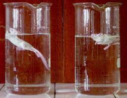
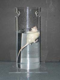
The behavioural despair test (or Porsolt forced swimming test) is a test, centred on a rodent’s response to the threat of drowning, whose result has been interpreted as measuring susceptibility to negative mood. It is commonly used to measure the effectiveness of antidepressants, although significant criticisms of its interpretation have been made.
An ‘interesting’ way to test the effectiveness of antidepressants!
Method
Animals are subjected to two trials during which they are forced to swim in an acrylic glass cylinder filled with water, and from which they cannot escape. The first trial lasts 15 minutes. Then, after 24-hours, a second trial is performed that lasts 5 minutes. The time that the test animal spends in the second trial without making any movements beyond those required to keep its head above water is measured. This immobility time is decreased by various types of antidepressants and also by electroconvulsive shock. Another common variant of this behavioural test specifically used for mice is conducted only for one trial and lasts six minutes. Modern implementations of the test score swimming and climbing behaviours separately, because swimming behaviour has been shown to be increased by selective serotonin reuptake inhibitors, while climbing behaviour is increased by selective norepinephrine reuptake inhibitors such as desipramine and maprotiline.
Controversy in Interpretation
Classically, immobility in the second test has been interpreted as a behavioural correlate of negative mood, representing a kind of hopelessness in the animal. Rodents given antidepressants swim harder and longer than controls (which forms the basis for claims of the test’s validity). However, there is some debate between scientists whether increased immobility instead demonstrates learning or habituation, and would therefore be a positive behavioural adaptation: the animal is less fearful because it is now familiar with the environment of the test. This interpretation is supported by the fact that even rats who are first put into a container from which they can escape (and therefore do not experience despair) show reduced mobility in the second test.
Some pharmacological compounds that influence motor movement, like stimulants and sedatives, may cause animals to swim for different amounts of time that are unrelated to the antidepressant properties of the compound. Researchers need to assess locomotor activity in the animal’s homecage or by a locomotor test. If locomotion is altered compared to controls then other animal antidepressant models should be used.
The term “behavioural despair test” bears an anthropomorphic connotation and is a somewhat subjective description as it is uncertain whether the test reliably gauges mood or despair. Strictly speaking, the descriptive term “forced swimming test” is preferred by researchers. The use of forced swimming tests is criticised by animal rights groups, notably PETA.
My Comments
I cannot believe some of the shi**y things researchers will do to animals.
Can you imagine being arbitrarily thrown into a container full of water, not knowing what is going on, not being able to touch the bottom, and no means of getting out of the container? Would you have a low mood or be in despair at your situation?
I see why they changed the name of the ‘test’, to sooth their conscience rather than that of the mice.
This page is based on the copyrighted Wikipedia article < https://en.wikipedia.org/wiki/Behavioural_despair_test >; it is used under the Creative Commons Attribution-ShareAlike 3.0 Unported License (CC-BY-SA). You may redistribute it, verbatim or modified, providing that you comply with the terms of the CC-BY-SA.
Azapirones have shown benefit in general anxiety and augmenting SSRIs in social anxiety and depression. Evidence is not clear for panic disorder and functional gastrointestinal disorders.
Tandospirone has also been used to augment antipsychotics in Japan as it improves cognitive and negative symptoms of schizophrenia. Buspirone is being investigated for this purpose as well.
Side Effects
Side effects of azapirones may include dizziness, headaches, restlessness, nausea, and diarrhoea.
Azapirones have more tolerable adverse effects than many other available anxiolytics, such as benzodiazepines or SSRIs. Unlike benzodiazepines, azapirones lack abuse potential and are not addictive, do not cause cognitive/memory impairment or sedation, and do not appear to induce appreciable tolerance or physical dependence. However, azapirones are considered less effective with slow onset in controlling symptoms.
Chemistry
Buspirone was originally classified as an azaspirodecanedione, shortened to azapirone or azaspirone due to the fact that its chemical structure contained this moiety, and other drugs with similar structures were labelled as such as well. However, despite all being called azapirones, not all of them actually contain the azapirodecanedione component, and most in fact do not or contain a variation of it. Additionally, many azapirones are also pyrimidinylpiperazines, though again this does not apply to them all.
Drugs classed as azapirones can be identified by their -spirone or -pirone suffix.
Pharmacology
Pharmacodynamics
On a pharmacological level, azapirones varyingly possess activity at the following receptors:
Actions at D4, 5-HT2C, 5-HT7, and sigma receptors have also been shown for some azapirones.
While some of the listed properties such as 5-HT2A and D2 blockade may be useful in certain indications such as in the treatment of schizophrenia (as with perospirone and tiospirone), all of them except 5-HT1A agonism are generally undesirable in anxiolytics and only contribute to side effects. As a result, further development has commenced to bring more selective of anxiolytic agents to the market. An example of this initiative is gepirone, which was recently approved after completing clinical trials in the United States for the treatment of major depression and generalized anxiety disorder. Another example is tandospirone which has been licensed in Japan for the treatment of anxiety and as an augmentation to antidepressants for depression.
5-HT1A receptor partial agonists have demonstrated efficacy against depression in rodent studies and human clinical trials. Unfortunately, however, their efficacy is limited and they are only relatively mild antidepressants. Instead of being used as monotherapy treatments, they are more commonly employed as augmentations to serotonergic antidepressants like the SSRIs. It has been proposed that high intrinsic activity at 5-HT1A postsynaptic receptors is necessary for maximal therapeutic benefits to come to prominence, and as a result, investigation has commenced in azapirones which act as 5-HT1A receptor full agonists such as alnespirone and eptapirone. Indeed, in preclinical studies, eptapirone produces robust antidepressant effects which surpass those of even high doses of imipramine and paroxetine.
Pharmacokinetics
Azapirones are poorly but nonetheless appreciably absorbed and have a rapid onset of action, but have only very short half-lives ranging from 1–3 hours. As a result, they must be administered 2-3 times a day. The only exception to this rule is umespirone, which has a very long duration with a single dose lasting as long as 23 hours. Unfortunately, umespirone has not been commercialized. Although never commercially produced, Bristol-Myers Squibb applied for a patent on 28 October 1993, and received the patent on 11 July 1995, for an extended release formulation of buspirone. An extended release formulation of gepirone is currently under development and if approved, should help to improve this issue.
Metabolism of azapirones occurs in the liver and they are excreted in urine and faeces. A common metabolite of several azapirones including buspirone, gepirone, ipsapirone, revospirone, and tandospirone is 1-(2-pyrimidinyl)piperazine (1-PP). 1-PP possesses 5-HT1A partial agonist and α2-adrenergic antagonist actions and likely contributes overall mostly to side effects.
This page is based on the copyrighted Wikipedia article < https://en.wikipedia.org/wiki/Azapirone >; it is used under the Creative Commons Attribution-ShareAlike 3.0 Unported License (CC-BY-SA). You may redistribute it, verbatim or modified, providing that you comply with the terms of the CC-BY-SA.


You must be logged in to post a comment.